
DNA origami
Automated Purification of DNA Origami
We have developed the application solid-phase reversible immobilization (SPRI) beads as a scalable, high-throughput, and automatable method to purify DNA origami .
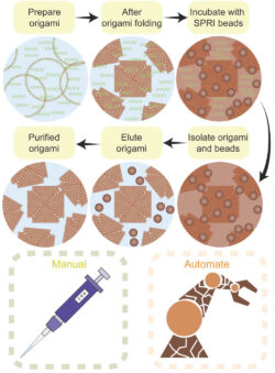
Figure 1. Schematic of the SPRI beads purification approach
This method remove unreacted oligos and biomolecules with yields comparable to existing methods while maintaining the high structural integrity of the DNA origami. It can also be integrated into an automated workflow to purify simultaneously large numbers and quantities of samples. We have worked with the Earlham Insitute to demonstrate the scalability of the approach and you can read more about the method in Small
Electrochemical Random Access DNA Memory (e-RADM)
We are developing a compartmentalized electrochemical random access DNA memory (e-RADM) using cascade reactions controlled by DNA nanostructures immobilized on gold microelectrode arrays. These nanostructures will be triggered when a specific information retrieval query is put into the system and microelectrodes containing the desired information can then be identified by Square Wave Voltammetry.
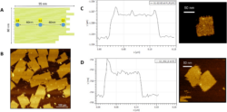
AFM images of the compartmentalised DNA origami
You can read more about the e-RADM approach in the MRS Advances paper
Supramolecular DNA nanostructures for single molecule sensing
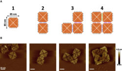
AFM images of supramolecular DNA nanostructures
We take advantage of the enhanced sensitivity of a nanopore that employs a poly-ethylene glycol enriched electrolyte to deliver real-time, non-destructive, and label-free fingerprinting of higher-order assemblies of DNA origami nanostructures with single-entity resolution. This approach enables the quantification of the assembly yields for complex DNA origami nanostructures . The nanopore readout provides analytical quantification of the complex supramolecular nanostructures within minutes, without any need for labelling and with single-molecule resolution.
You can read more about this work in the Biophysical Journal paper
Visualizing and Quantifying microRNA-Induced DNA Origami Separation at the Nanoscale
Clinically relevant miRNA biomarkers trigger the disassembly of DNA origami dimers into monomers through a toehold-mediated strand displacement reaction. High-speed AFM was used to visualize this reaction in real time, while solid-state nanopore measurements quantified the populations of dimers and monomers, as well as the resulting miRNA concentration, even in an RNase-contaminated environment. This robust, near-irreversible process enables multiplexed miRNA detection in the presence of RNases, offering a new and reliable route for small RNA sensing.

Localization AFM images of the linkers connecting DNA origami dimers.
You can read more about this work in the Angewandte Chemie paper
Work with us
We love collaborating on exciting scientific projects. If you are interested about working with, contact Prof Christoph Wälti...We would love to hear from you!!!