Research
Biosensing
Enzyme-Switches
We have used and developed enzyme-switch assays for quick, easy, and sensitive analyte measurement. Traditional immunoassays require a number of wash steps, necessitating manual handling or complex automation systems. Our developed enzyme-switches are wash free, enabling simple analyte measurement for use in a wide range of settings, including low-resource settings and at the point-of-care.
The "BLA-BLIP" assay was first developed in 2013 by Maarten Merkx group (ACS Chem. Biol. 2013, 8, 10, 2127–2132). It utilises TEM-1β-lactamase (BLA) and its inhibitor β-lactamase inhibitor protein (BLIP), which are tethered together with a long flexible linker containing two biorecognition elements. We have incorporated Affimers as biorecognition elements in the BLA-BLIP system for the detection of a number of analytes. Affimers are non-immunoglobulin-binding proteins and offer the benefit of being small, stable, and easily expressed as recombinant proteins when fused genetically to split-enzyme fragments.

The NanoBit enzyme-switch sensor combines Affimer proteins as biorecognition elements with a split-luciferase, NanoLuc® Binary Technology (NanoBiT, Promega). The NanoBit system relies on the NanoLuc split-luciferase enzyme (ACS Chem. Biol. 2016, 11, 2, 400–408). We have recombinantly fused Affimer biorecognition elements to the LgBit and the SmBit, respectively, via a semi-flexible peptide linker (Figure 2).

Therapeutic Drug Monitoring (TDM)
Therapeutic drug monitoring (TDM) is the practice of measuring drug concentrations in patient samples and adjusting dosing towards an optimal concentration, to personalise care. TDM is a complicated intervention that requires taking blood samples at specified points across the therapy, accurately measuring the active drug in the sample, calculating the amount of drug to give the patient in the next dose, and reporting results and recommendations to clinicians. Currently, TDM is challenging as standard techniques for measuring concentrations, particularly for small molecule drugs, can be prohibitive for TDM. Liquid chromatography coupled mass spectrometry (HPLC-MS/MS) is the gold-standard for small molecule measurement. HPLC-MS/MS requires specialised facilities and staff, it is expensive and introduces more logistical challenges in sending samples to centralised laboratories. We are working towards the implementation of our biosensor technology for quick and accessible TDM for a wide range of clinical settings, including at the point-of-care.
DNA origami
Automated Purification of DNA Origami
We have developed the application solid-phase reversible immobilization (SPRI) beads as a scalable, high-throughput, and automatable method to purify DNA origami .
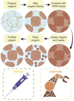
Figure 1. Schematic of the SPRI beads purification approach
This method remove unreacted oligos and biomolecules with yields comparable to existing methods while maintaining the high structural integrity of the DNA origami. It can also be integrated into an automated workflow to purify simultaneously large numbers and quantities of samples. We have worked with the Earlham Insitute to demonstrate the scalability of the approach and you can read more about the method in Small
Electrochemical Random Access DNA Memory (e-RADM)
We are developing a compartmentalized electrochemical random access DNA memory (e-RADM) using cascade reactions controlled by DNA nanostructures immobilized on gold microelectrode arrays. These nanostructures will be triggered when a specific information retrieval query is put into the system and microelectrodes containing the desired information can then be identified by Square Wave Voltammetry.
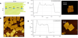
Figure 1. AFM images of the compartmentalised DNA origami
You can read more about the e-RADM approach in the MRS Advances paper
Supramolecular DNA nanostructures for single molecule sensing
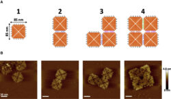
Figure 3. AFM images of supramolecular DNA nanostructures
We take advantage of the enhanced sensitivity of a nanopore that employs a poly-ethylene glycol enriched electrolyte to deliver real-time, non-destructive, and label-free fingerprinting of higher-order assemblies of DNA origami nanostructures with single-entity resolution. This approach enables the quantification of the assembly yields for complex DNA origami nanostructures . The nanopore readout provides analytical quantification of the complex supramolecular nanostructures within minutes, without any need for labelling and with single-molecule resolution.
You can read more about this work in the Biophysical Journal paper
Nanopore
Our research aims to develop new tools for single-cell analysis and manipulation with single molecule resolution to study the dynamic function of individual cells in heterogeneous populations.
Single Cell Analysis
Single-cell Nanosurgery. We are developin an electrical nanobiopsy platform capable of extracting genetic material and organelles from single cells in culture. We are applying this technology to understand why a brain cancer called GBM is so deadly and what we can do to stop it. In 2024, we published a paper in Science Advances describing longitudinal transcriptomics of cancer cells . You can also read here the University of Leeds press release on our work.
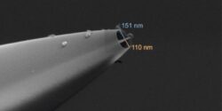
Figure 1. SEM micrographs of a double-barrel nanopipette for single cell nanosurgery
Single-molecule Nanoinjection. We are developing a platform that can inject biomolecules one at the time into living cells in culture. We are working with Dr Eric Hewitt and Prof Sheena Radford OBE, FMedSci, FRS to understand the mechanism of neurodegeneration in Parkinson’s disease.

Figure 2. Artistic illustration of SICM micrographs of living cells
Read our Nature Communications paper describing single molecule delivery into living cells.
Nanopore Sensing
Single molecule Biosensing. We are developing functionalized DNA origami nanostructures to capture disease biomarkers within biological fluids. We can analyse these nanostructures one at the time using nanopore sensors aiming to detect the tiniest amount of disease biomarkers so that a diagnosis can be done as quickly as possible.
You can read more about our work in our Nature Communications paper on the rational design of DNA origami nanostructures and in our Biophysical Journal article describing fingerprinting of supramolecular DNA nanostructures.

Figure 3. Schematic of the DNA origami-nanopore biosensing approach
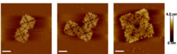
Figure 4. AFM micrographs Supramolecular DNA origami nanostructures
Organs-on-chip
We are generally interested in the development of the Organ-on-a-chip technology (OOC) and improving its adoption in the drug discovery process and for scientific research and for this reason we are keen to evaluate improvements and challenges to its adoption (link).
With collaborators in many different institutes, we help with the optimization of the organ-on-a-chip to be able to model different physiological mechanisms. Here some examples:
- Neurovascular Unit on a Chip
In my publication Recreating Blood-Brain Barrier Physiology and Structure on Chip: A Novel Neurovascular Microfluidic Bioreactor, I contributed to the design and optimization of one of the first microfluidic device that mimics the human blood-brain barrier. This platform helps researchers study neurovascular functions and disorders. - Placenta-on-a-Chip Models
I have contributed to the development of on-a-chip models of the placenta and the fetal membranes which allow us to study maternal-fetal interactions, drug toxicity and bacterial infection during pregnancy. These models provide critical insights into placental function and pregnancy-related diseases.
Our main focus however remain the development of enabling technologies to improve our understanding of human reproduction by introducing physiologically relevant in vitro models, and developing new platforms and processes to improve fertility treatment.
- Organs-on-a-Chip Models of the Female Reproductive System
In this comprehensive review, Organs-On-Chip Models of the Female Reproductive System, we analysed microfluidic-based technologies that mimic in vivo conditions of female reproductive organs. These models hold great promise for studying diseases and pregnancy-related complications such as pre-eclampsia and infertility. - Endometrium-on-a-chip The endometrial environment is complex, dynamically changing and its functioning mechanisms are finely tuned to support women health as well as the establishment of a successful pregnancy. The factors that influence these mechanisms are not completely understood and most of this is due to the complexity to model them in vitro. With our general approach we collaborate with several groups to develop new models based on microfluidics, organoids and more.
- Microfluidic Devices for Embryo Culture
I led the project Design, Fabrication, and Testing of a Mouse Embryo Culture Chip, funded by the NC3Rs CRACK-IT Challenge. This initiative aimed to create microfluidic devices that enhance embryo handling and culture, improving assisted reproductive technologies while also reducing reliance on animal models. This technology has been now translated into a commercial device with the University of Leeds' Spin off IVFmicro ltd.
Work with us
We love collaborating on exciting scientific projects. If you are interested about working with us, contact Prof Christoph Wälti, Dr Virginia Pensabene or Dr Paolo Actis...we would love to hear from you!!!